
BARTON VS COLLES FRACTURE SKIN
Open wound and/or the bone protruding through the skin (open fractures require different management due to the risk of infection). Less common clinical findings may include: Tenderness on palpation of the distal radius. Pallor: pallor or duskiness of the hand (suggesting vascular injury)Ī thorough orthopaedic examination of the hand and wrist, along with a neurological examination of the upper limb should be performed to assess functional capacity and neurovascular deficits. Pain: disproportionate to the injury (suggesting compartment syndrome). Paraesthesia: tingling, pins and needles or loss of sensation in the hand (suggesting nerve injury). Social history: smoking and alcohol (risk factors for osteoporosis), occupation (may influence the choice of management)įor more information on falls, see the Geeky Medics guide to the assessment, investigation and management of falls.Īlthough less common, it is important to ask about symptoms which may suggest neurovascular injury including the 3P’s:. Family history: a family history of fragility fractures, especially parental hip fractures, is a risk factor for osteoporosis. Past medical history: risk factors for osteoporosis, previous fragility fractures, co-morbidities. Clinical features of neurovascular compromise (see below). Events surrounding the fall: features suggestive of syncope, other injuries (e.g. Other important areas to cover in the history include: Typical symptoms of distal radius fractures include: The mechanism of injury and clinical findings, including skin integrity, assessment of circulation and sensation, should be documented at presentation. Parkinson’s, disease, stroke)Ī thorough history and examination of the injury should be performed for patients who present with a suspected distal radius fracture. 
An overview of risk factors for fractures of the distal radius.

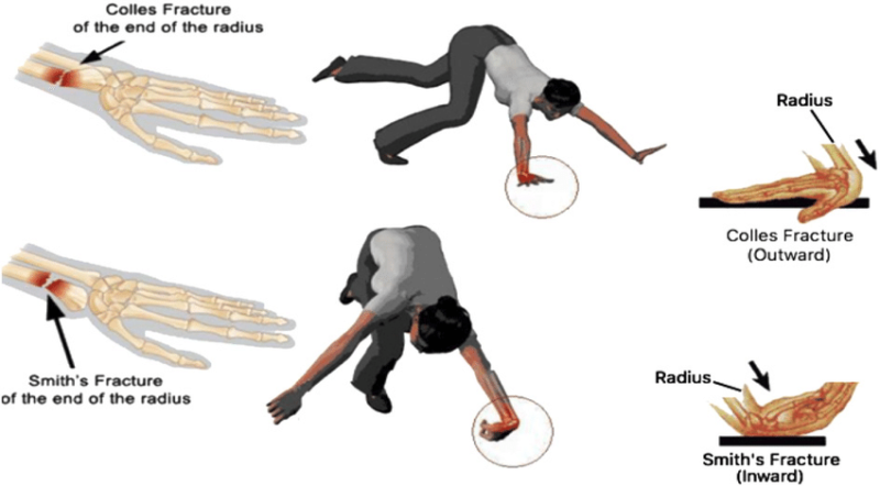
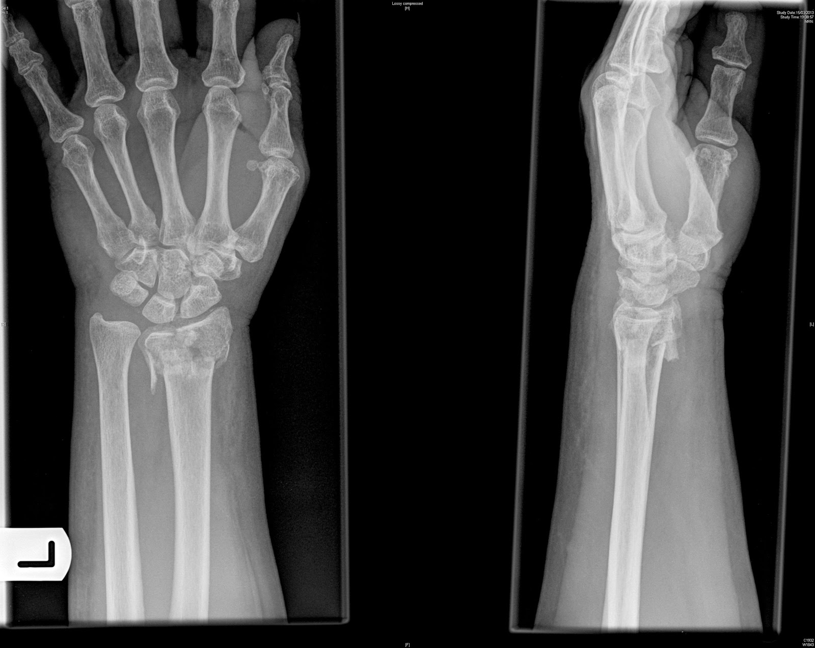
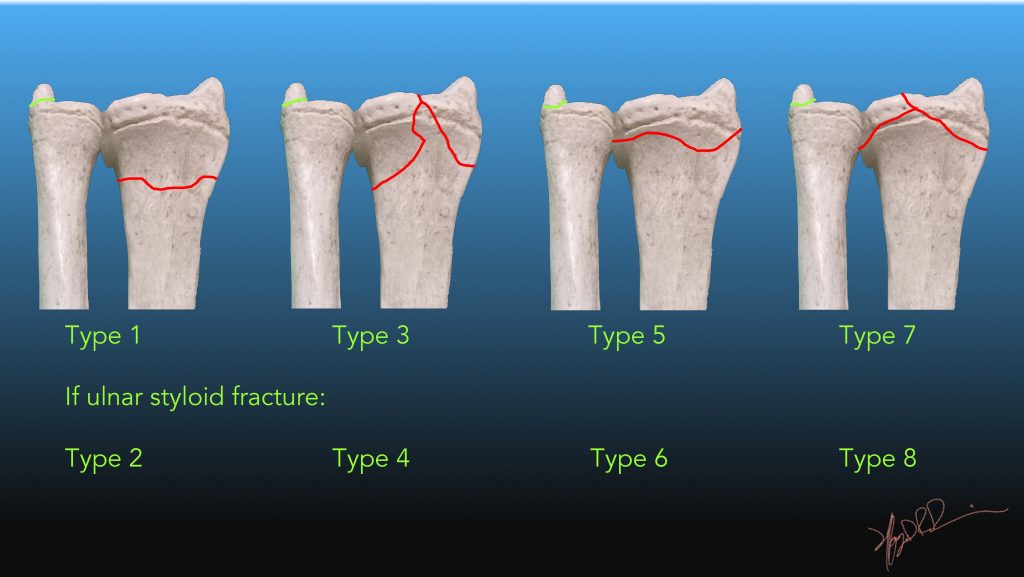
Risk factors can be thought of in terms of those which increase the risk of osteoporosis and those which increase the risk of falling (Table 1). Barton’s fracture: an intra-articular fracture with associated dislocation of the radiocarpal joint.Smith’s fracture: an extra-articular fracture with volar displacement.Colles’ fracture (most common): an extra-articular fracture with dorsal displacement (“dinner fork deformity”).There are three key fractures of the distal radius to be aware of: blood tests including FBC, serum calcium, alkaline phosphatase and an isotope bone scan). If atraumatic, a pathological fracture should be suspected, and further workup should investigate for malignancy (e.g. The most common mechanism of injury is a fall onto an outstretched hand ( FOOSH). Elderly patients with osteoporosis and low energy trauma.Young patients who participate in sport or are involved in trauma.Aetiologyįractures of the distal radius are most common in two main groups of patients: 3 The Frykman classification of distal radial fractures is based on the AP appearance and encompasses the eponymous entities of Colles fracture, Smith fracture, Barton fracture, chauffeur fracture. It assesses the pattern of fractures, involvement of the radioulnar joint and presence of a distal ulnar fracture.You might also be interested in our surgical flashcard collection which contains over 500 flashcards that cover key surgical topics.


 0 kommentar(er)
